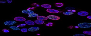
Fluorescence Microscopy
Microscopy is at the heart of understanding tissue architecture, cell structure, and dynamics, as well
as molecular function. Fluorescence microscope" refers to any microscope that uses fluorescence to
generate an image. In this case, a simple setup uses epifluorescence to generate images with a
combination of filters and dichroic mirrors.
- Culture of cells
- Treatment and transfection of cells
- Immunostaining
- Imaging of the cells
Cell Imaging
Modern biology relies heavily on cell imaging, which enables scientists to see and examine the dynamics,
structure, and function of cells in great detail. To investigate cellular processes in fixed or live
samples, it includes a number of methods such as fluorescence microscopy, live-cell imaging, and
high-content screening.
-
Brightfield Microscopy
- Phase-Contrast Microscopy
- Fluroscence Microscopy
- Live-Cell Imagin
g
.
Karyotyping Analysis
Karyotyping is the process of viewing chromosomes under a microscope during the metaphase phase of cell
division and organizing them into a karyogram using a standard format that includes banding patterns,
size, and shape.
- Chromosomal Abnormalities
- Prenatal Testing
- Infertility and Miscarriage Analysis
- Cancer Diagnosis
- Reproductive Medicine
.
Fluorescence In-Situ Hybridization (FISH)
A molecular cytogenetic approach called fluorescence in situ hybridization (FISH) locates and
identifies particular DNA sequences on chromosomes or inside cells using fluorescent probes. Because
of its accuracy and capacity to identify particular genetic disorders, this method is frequently used
in clinical diagnostics, genetic research, and oncology.
CONTACT
:
Email:
info@allelelifesciences.com or
Whatsapp
+91- 8377082003