

Cell and Tissue Structure Visualization.
- Read More
Immunofluorescence microscopy is a
powerful technique used to visualize the
presence and location of specific
proteins or antigens in biological
samples using fluorescently labeled
antibodies. It's widely used in cell
biology, pathology, and diagnostic
medicine.
Services:
» Direct
Immunofluorescence
» Indirect
Immunofluorescence
» Live-Cell
Immunofluorescence
» DAPI or Hoechst
stains to visualize nuclei
» SYBR®
Green or FITC Staining
Immunofluorescence microscopy depicts the full distribution of the epitope that the antibody recognizes and analyzes steady state localization.


These labeled
antibodies bind directly or indirectly
to cellular antigens. The technique has
a number of different biological
applications including evaluation of
cells in suspension, cultured cells and
tissue.
- Infectious Disease Diagnosis
- Autoimmune Diseases
- Cancer Diagnostics and Research
- Genetic Disorders
- Neurological Disorders
The fluorescence
can be visualized using fluorescence
microscopy. The IF technique allows for
a visualization of the presence as well
as the distribution of target molecules
in a sample.
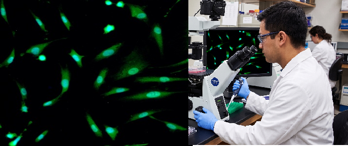
Note: An illustrative fluorescence microscopy field showing labeled cells. Your delivered images will show your specific antibody/marker staining pattern at full resolution. The chart above is illustrative artwork for orientation only and does not represent actual client data.
Immunofluorescence microscopy is a powerful technique used to visualize the presence and location of specific proteins or antigens in biological samples using fluorescently labeled antibodies. It's widely used in cell biology, pathology, and diagnostic medicine. Services :
These labeled antibodies bind directly or indirectly to cellular antigens. The technique has a number of different biological applications including evaluation of cells in suspension, cultured cells and tissue.